Pelvic Anatomy Xray / Ortho Dx A Woman With A Hip That Is Popping Out Of Place Clinical Advisor - Branches of the internal iliac artery.. Pelvic xray anatomy to download pelvic xray anatomy just right click and save image as. The pelvic diaphragm is composed of the ischiococcygeus muscle and levator ani muscle, the latter of which consists of the iliococcygeus, puborectalis, and pubococcygeus muscles. Pelvic_xray_anatomy.png (596 × 527 pixels, file size: In an adult, the innominate bones consist of the fused ilium, ischium, and pubis (figure 1). Surgical pelvic anatomy in gynecologic oncology.
Our latest youtube film is ready to run. Pelvic anatomy mri variant anatomy pelvic viscera. The cortex of femoral head, neck, greater, and lesser trochanter should be smooth with normal trabecular pattern on ap and lateral. Use the mouse scroll wheel to move the images up and down alternatively use the tiny arrows (>>) on both side of the image to move the images.>>) on both side of the image to move the images. Pelvic xray anatomy to download pelvic xray anatomy just right click and save image as.
Normal Pelvis X Ray Ap Radiology Case Radiopaedia Org from prod-images-static.radiopaedia.org Our latest youtube film is ready to run. 6.1a, b) is a bony ring consisting of paired innominate bones, the sacrum and coccyx. Annotated case courtesy of dr phillip marsh, radiopaedia.org, rid: Pelvis anatomy the pelvis is either the lower part of the trunk of the human body between the abdomen and the thighs. Magnetic resonance imaging or mri of the female pelvis offers a unique display of the pelvic anatomy, including a woman's ovaries, uterus, and fallopian tubes. There is a printable worksheet available for download here so you can take the quiz with. Mri of the female pelvis: Pelvic ring formed from 2 innominate.
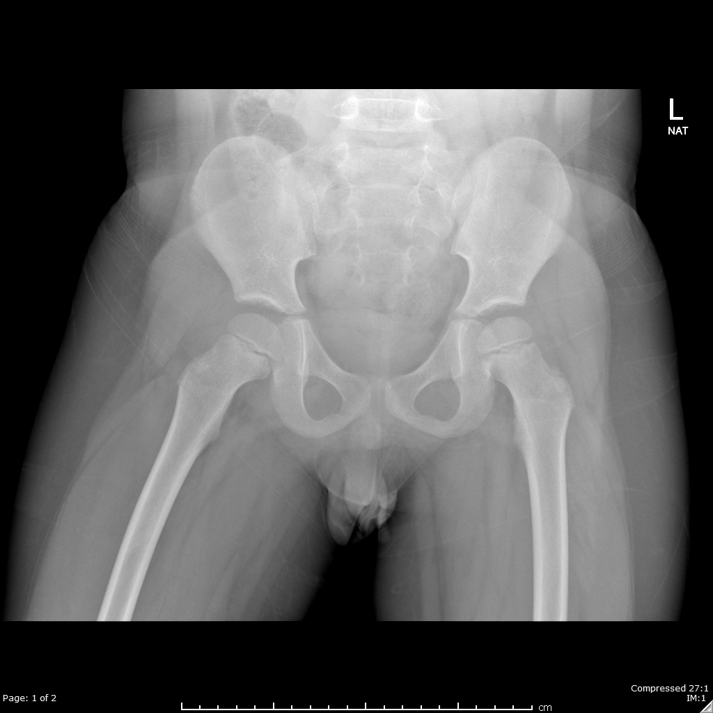
Pelvis x ray anatomy in this image you will find the sacroiliac joint acetabular obturator foramina greater trochanter pubic symphysis femoral heads lesser trochanters in it.
Each innominate bone is composed of three parts, which fuse at the acetabulum. Anatomy of ilioinguinal and iliohypogastric nerves in relation to trocar placement and low transverse incisions. Pelvis anatomy the pelvis is either the lower part of the trunk of the human body between the abdomen and the thighs. Pelvic anatomy mri variant anatomy pelvic viscera. Mands thorough break down of this commonly used ed diagnostic the pelvic xr. 6.1a, b) is a bony ring consisting of paired innominate bones, the sacrum and coccyx. Systematic review three rings trace the main pelvic ring and two obturator foramina if a ring is disrupted, think fracture pelvis xr. Pelvic anatomy on mri ashish p. To review pelvic sidewall anatomy including retroperitoneal spaces. Hemi pelvis anatomy normal ap. It is subdivided into the greater pelvis and lesser pelvis. In an adult, the innominate bones consist of the fused ilium, ischium, and pubis (figure 1). An x ray of the pelvis focuses specifically on the area between your hips that holds many of your reproductive.
Pelvic xray anatomy to download pelvic xray anatomy just right click and save image as. Branches into common femoral artery (distal to inguinal ligament) An x ray of the pelvis focuses specifically on the area between your hips that holds many of your reproductive. This mri male pelvis axial cross sectional anatomy tool is absolutely free to use. Magnetic resonance imaging or mri of the female pelvis offers a unique display of the pelvic anatomy, including a woman's ovaries, uterus, and fallopian tubes.
Normal Pelvis X Ray 4 Year Old Radiology Case Radiopaedia Org from prod-images-static.radiopaedia.org Pelvis x ray anatomy in this image you will find the sacroiliac joint acetabular obturator foramina greater trochanter pubic symphysis femoral heads lesser trochanters in it. The main purposes of the pelvic girdle are to support and protect the abdominal and pelvic organs, and to connect the trunk and lower limbs. Axial, coronal, sagittal, and 3d reconstructions accompany highly accurate and detailed medical drawings, assisting you in making an accurate diagnosis. Pelvis anatomy the pelvis is either the lower part of the trunk of the human body between the abdomen and the thighs. Siu/icud consultation on urethral strictures: If cortical disruption, trabecular pattern disruption or transverse sclerosis, think fractured proximal femur. Hover on/off image to show/hide findings. Branches of the internal iliac artery.
The cortex of femoral head, neck, greater, and lesser trochanter should be smooth with normal trabecular pattern on ap and lateral.
Pelvic ring formed from 2 innominate. Chest, abdomen, pelvis provides detailed views of anatomic structures in successive imaging slices in each standard plane of imaging. Surgical pelvic anatomy in gynecologic oncology. Hemi pelvis anatomy normal ap. Articulate posteriorly with the sacrum and anteriorly through pubis symphysis. ●to review pelvic sidewall anatomy including retroperitoneal spaces. It includes several structures : Pelvic anatomy on mri ashish p. Hemi pelvis anatomy normal ap. The bony pelvis, the pelvic cavity, the pelvic floor, and the perineum. If line disruption, think fractured proximal femur. Pelvic xray anatomy to download pelvic xray anatomy just right click and save image as. Each innominate bone is composed of three parts, which fuse at the acetabulum.
An x ray of the pelvis focuses specifically on the area between your hips that holds many of your reproductive. Articulate posteriorly with the sacrum and anteriorly through pubis symphysis. Pelvic_xray_anatomy.png (596 × 527 pixels, file size: The space or compartment surrounded by the pelvic girdle (bony pelvis). Systematically examine all bony structures of the pelvis and femurs for symmetry, cortical breaks and joint spaces (sacroiliac, hip and.
Radiography Pelvis Technique In Cats Vetlexicon Felis From Vetlexicon Definitive Veterinary Intelligence from www.vetstream.com Angiography invasive angiography is the gold standard modality for assessing pelvic vasculature 3. The innominate bones articulate with each other anteriorly and with the sacrum posteriorly. This mri male pelvis axial cross sectional anatomy tool is absolutely free to use. Anatomy pelvis at university of kansas medical center the sacroiliac joints should be symmetrical joint space range 2 4 mm. Hemi pelvis anatomy normal ap. Learn vocabulary, terms and more with flashcards only rub 220.84/month. An x ray of the pelvis focuses specifically on the area between your hips that holds many of your reproductive. The space or compartment surrounded by the pelvic girdle (bony pelvis).
Annotated case courtesy of dr matthew lukies, radiopaedia.org, 51247.
Mands thorough break down of this commonly used ed diagnostic the pelvic xr. Branches of the internal iliac artery. Click image to align with top of page. Hemi pelvis anatomy normal ap. Pelvic anatomy knowledge, and on participant confidence with imaging in clinical situations. There is a printable worksheet available for download here so you can take the quiz with. To review pelvic sidewall anatomy including retroperitoneal spaces. Pelvis anatomy the pelvis is either the lower part of the trunk of the human body between the abdomen and the thighs. Mri of the female pelvis: Anatomy of the pelvis the pelvis is a ring of bones situated between the spine and the legs. Representative images of normal pelvic anatomy, with select videos. Figure 3a schematics show the anatomy of the female pelvic floor at the level of the pelvic diaphragm (a) and the urogenital diaphragm (b). The muscle originates from the body of the pubis and attaches to the pectineal line and proximal part of the linea aspera of femur.


0 Komentar